Research: Neutron Radiography/Imaging
One of the key applications of neutron generators is imaging, and some of Adelphi’s customers use our generators for imaging Applications. Occasionally, we have been able to perform some imaging measurements either in-house at Adelphi Technology, and sometimes we have been able to run measurements at customers's sites or at the labs of government labs when we have participated on collaboratives research programs.
Adelphi Technology has published the following papers on imaging.
Williams, David L., Craig M. Brown, David Tong, Alexander Sulyman, and Charles K. Gary.
"A fast neutron radiography system using a high yield portable DT neutron source."
Journal of imaging 6, no. 12 (2020): 128.
Resolution measurements were made using 14.1 MeV neutrons from a high-yield, portable DT neutron generator and a neutron camera based on a scintillation screen viewed by a digital camera. Resolution measurements were made using a custom-built, plastic, USAF-1951 resolution chart, of dimensions 125 × 98 × 25.4 mm3, and by calculating the modulation transfer function from the edge-spread function from edges of plastic and steel objects. A portable neutron generator with a yield of 3 × 10⁹ n/s (DT) and a spot size of 1.5 mm was used to irradiate the object with neutrons for 10 min. The neutron camera, based on a 6LiF/ZnS:Cu-doped polypropylene scintillation screen and digital camera was placed at a distance of 140 cm, and produced an image with a spatial resolution of 0.35 cycles per millimeter.
Bergaoui, K., N. Reguigui, C. K. Gary, J. T. Cremer, J. H. Vainionpaa, and M. A. Piestrup.
"Design, testing and optimization of a neutron radiography system based on a
Deuterium–Deuterium
(D–D)
neutron generator."
Journal of Radioanalytical and Nuclear Chemistry 299 (2014): 41-51.
Simulations show that significant improvement in imaging performance can be achieved through collimator design for thermal and fast neutron radiography with a laboratory neutron generator. The radiography facility used in the measurements and simulations employs a fully high-voltage-shielded, axial D–D neutron generator with a radio frequency driven ion source. The maximum yield of such generators is about 1010 fast neutrons per seconds (E = 2.45 MeV). Both fast and thermal neutron images were acquired with the generator and a Charge Coupled Devices camera. To shorten the imaging time and decrease the noise from gamma radiation, various collimator designs were proposed and simulated using Monte Carlo N-Particle Transport Code (MCNPX 2.7.0). Design considerations included the choice of material, thickness, position and aperture for the collimator. The simulation results and optimal configurations are presented.
Cremer, J. T., D. L. Williams, C. K. Gary, M. A. Piestrup, D. R. Faber, M. J. Fuller, J. H.
Vainionpaa,
M.
Apodaca, R. H. Pantell, and J. Feinstein.
"Large area imaging of hydrogenous materials using fast neutrons from a DD fusion
generator."
Nuclear Instruments and Methods in Physics Research Section A: Accelerators,
Spectrometers,
Detectors and
Associated Equipment 675 (2012): 51-55.
A small-laboratory fast-neutron generator and a large area detector were used to image hydrogen-bearing materials. The overall image resolution of 2.5 mm was determined by a knife-edge measurement. Contact images of objects were obtained in 5–50 min exposures by placing them close to a plastic scintillator at distances of 1.5 to 3.2 m from the neutron source. The generator produces 109 n/s from the DD fusion reaction at a small target. The combination of the DD-fusion generator and electronic camera permits both small laboratory and field-portable imaging of hydrogen-rich materials embedded in high density materials.
https://www.sciencedirect.com/science/article/pii/S0168900212001386
Cremer, J. T., M. A. Piestrup, H. Park, C. K. Gary, R. H. Pantell, C. J. Glinka, and J. G.
Barker.
"Imaging hydrogenous materials with a neutron microscope."
Applied Physics Letters 87, no. 16 (2005).
Magnified images of materials containing hydrogen, for which the main contrast mechanism for neutrons is incoherent scattering, have been obtained using a microscope employing a neutron compound refractive lens (CRL). The CRL was composed of 100 biconcave lenses that produced magnified images of polyethylene and polypropylene (hydrogen-rich) grids and biological specimens using Å cold neutrons with a 10% bandwidth. For hydrogenous materials, 98%–99% of the attenuation is by incoherent scattering and 1%–2% from neutron absorption by the hydrogen nuclei. The small angle of acceptance of the CRL discriminates against scattered neutrons from the hydrogenous object, thereby producing the needed contrast for imaging.
Cremer, Jay Theodore, Melvin A. Piestrup, and Xizeng Wu.
"Fast and thermal neutron radiography."
Penetrating Radiation Systems and Applications VII, vol. 5923, pp. 46-64. SPIE, 2005.
here is a need for high brightness neutron sources that are portable, relatively inexpensive, and capable of neutron radiography in short imaging times. Fast and thermal neutron radiography is as an excellent method to penetrate high-density, high-Z objects, thick objects and image its interior contents, especially hydrogen-based materials. In this paper we model the expected imaging performance characteristics and limitations of fast and thermal radiography systems employing a Rose Model based transfer analysis. For fast neutron detection plastic fiber array scintllators or liquid scintillator filled capillary arrays are employed for fast neutron detection, and 6Li doped ZnS(Cu) phosphors are employed for thermal neutron detection. These simulations can provide guidance in the design, construction, and testing of neutron imaging systems. In particular we determined for a range of slab thickness, the range of thicknesses of embedded cracks (air-filled or filled with material such as water) which can be detected and imaged.
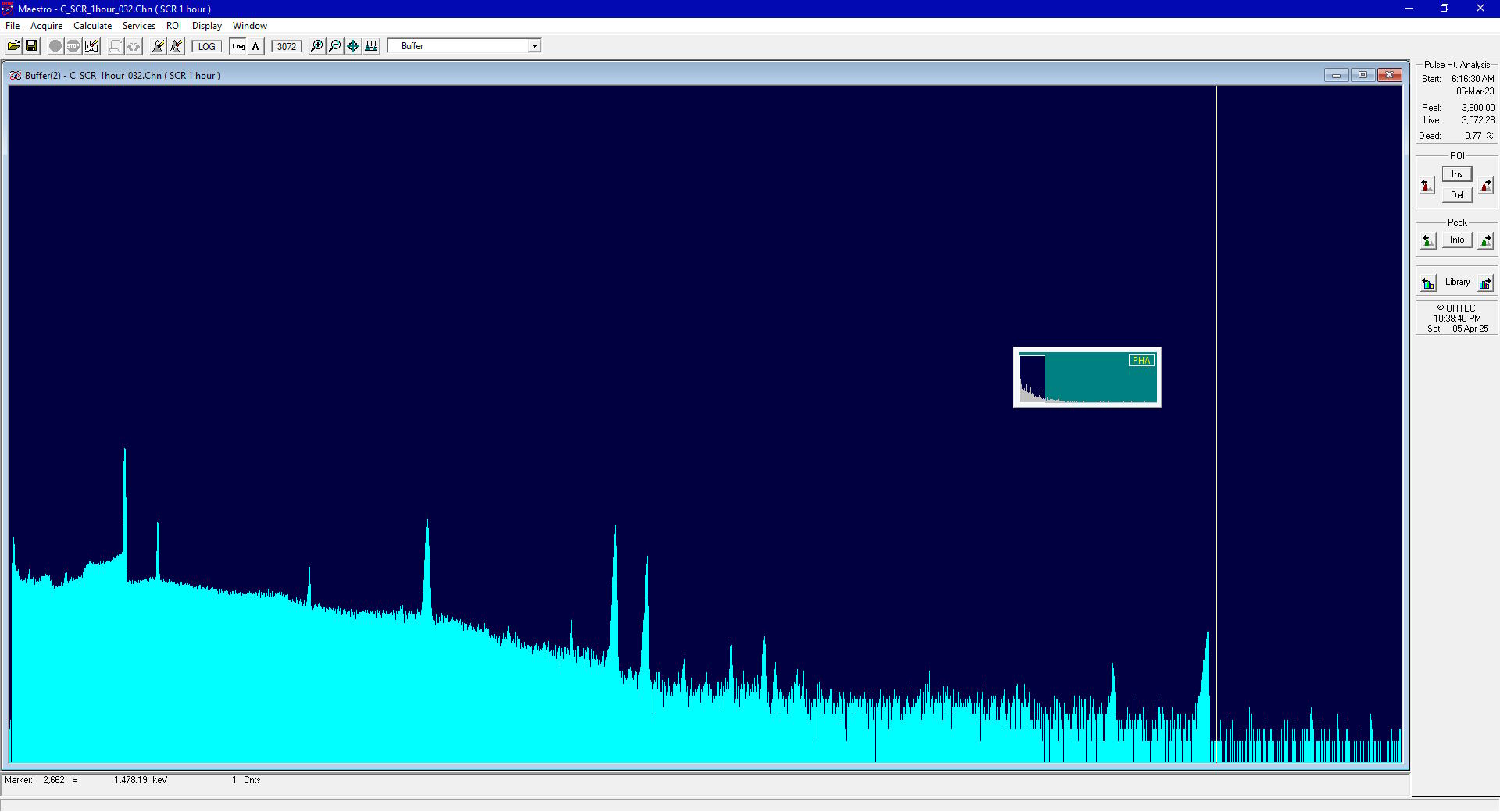
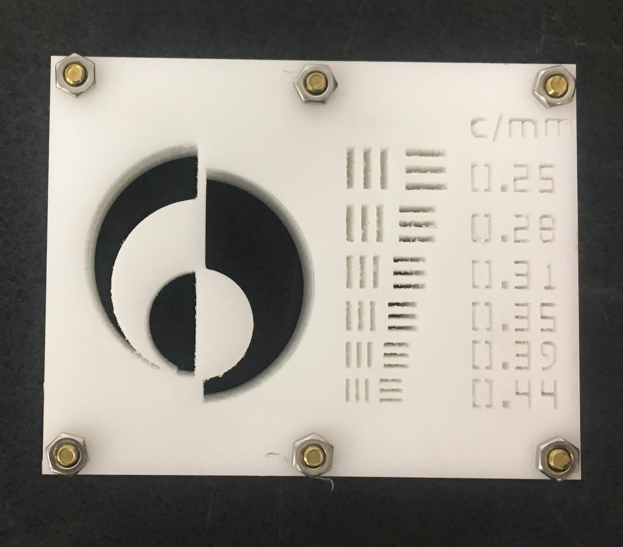
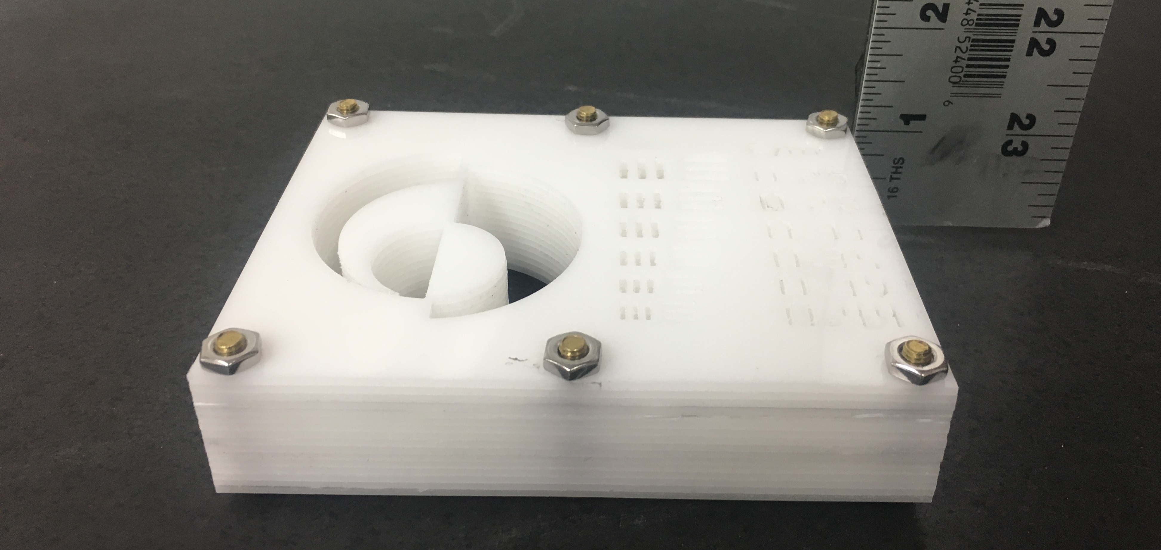
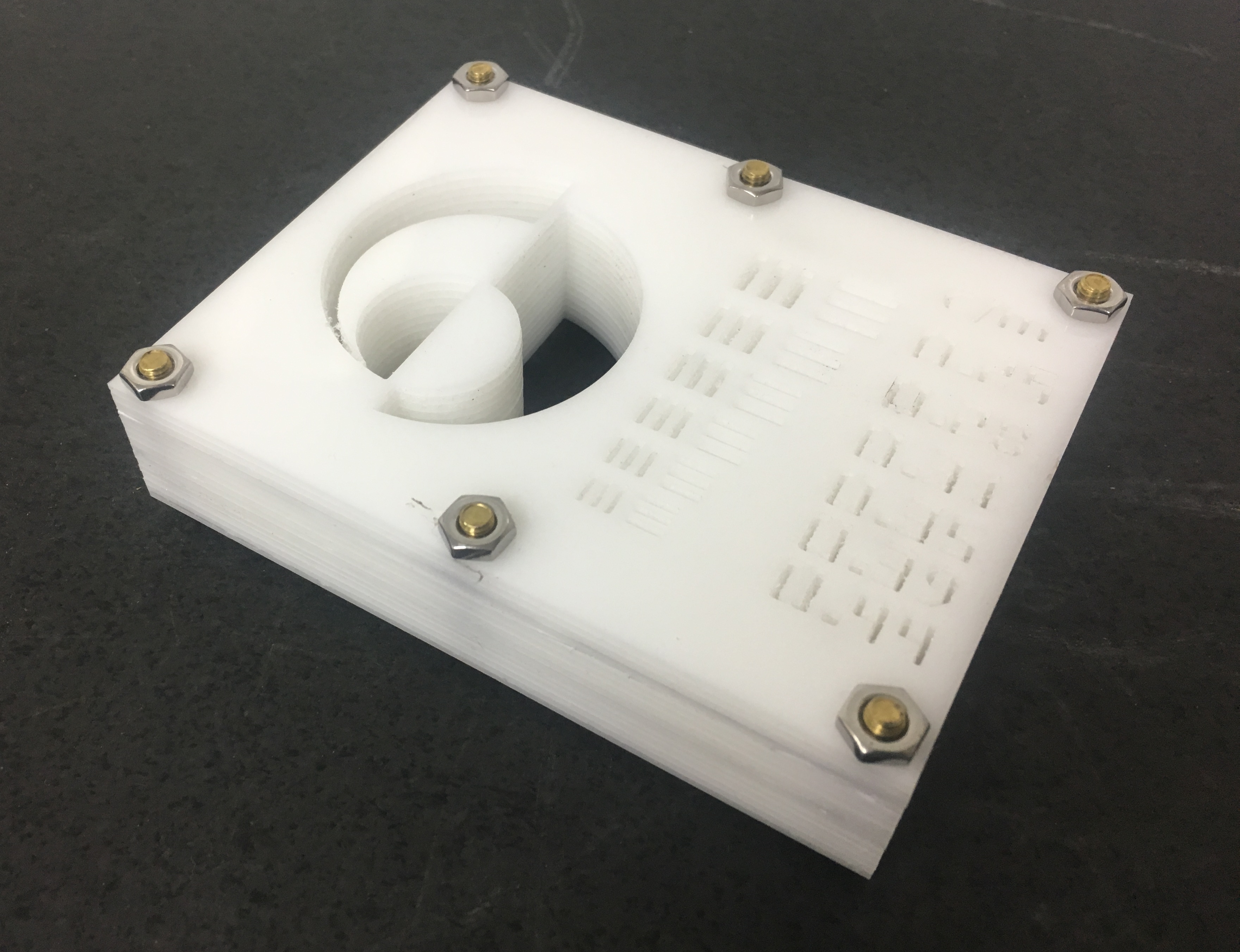
Resolution measurements have been made using the approaches of measuring an AF1951 imaging phantom (otherwise known as an imaging standard or target), a "Siemens Star" and by using the Modultion Transfer Function (MTF) technique.
Work on Fast Neutron Radiography (FNR) has included 14.1 MeV and 2.45 MeV imaging as well as Thermal Neutron Radiography (TNR). Some neutron radiograph images are given below.
Images
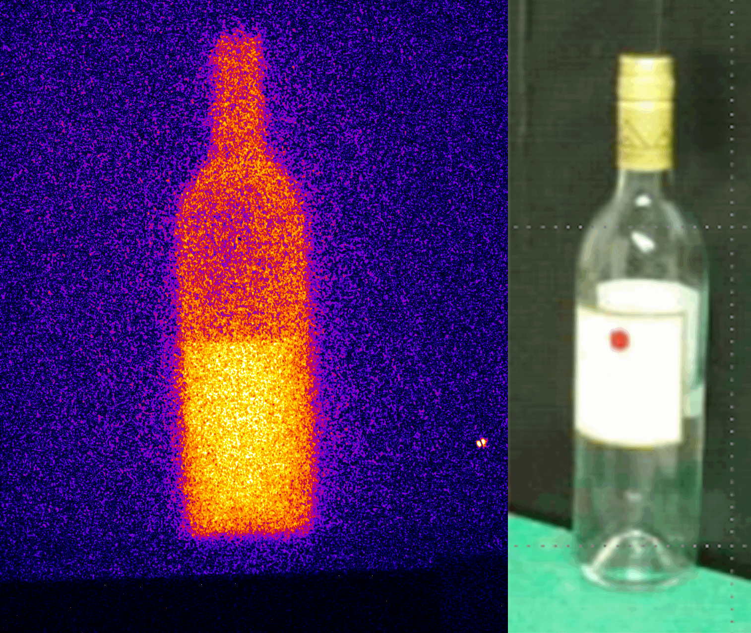
Wine bottle
2.45 MeV neutron imaging, winebottle (2008)
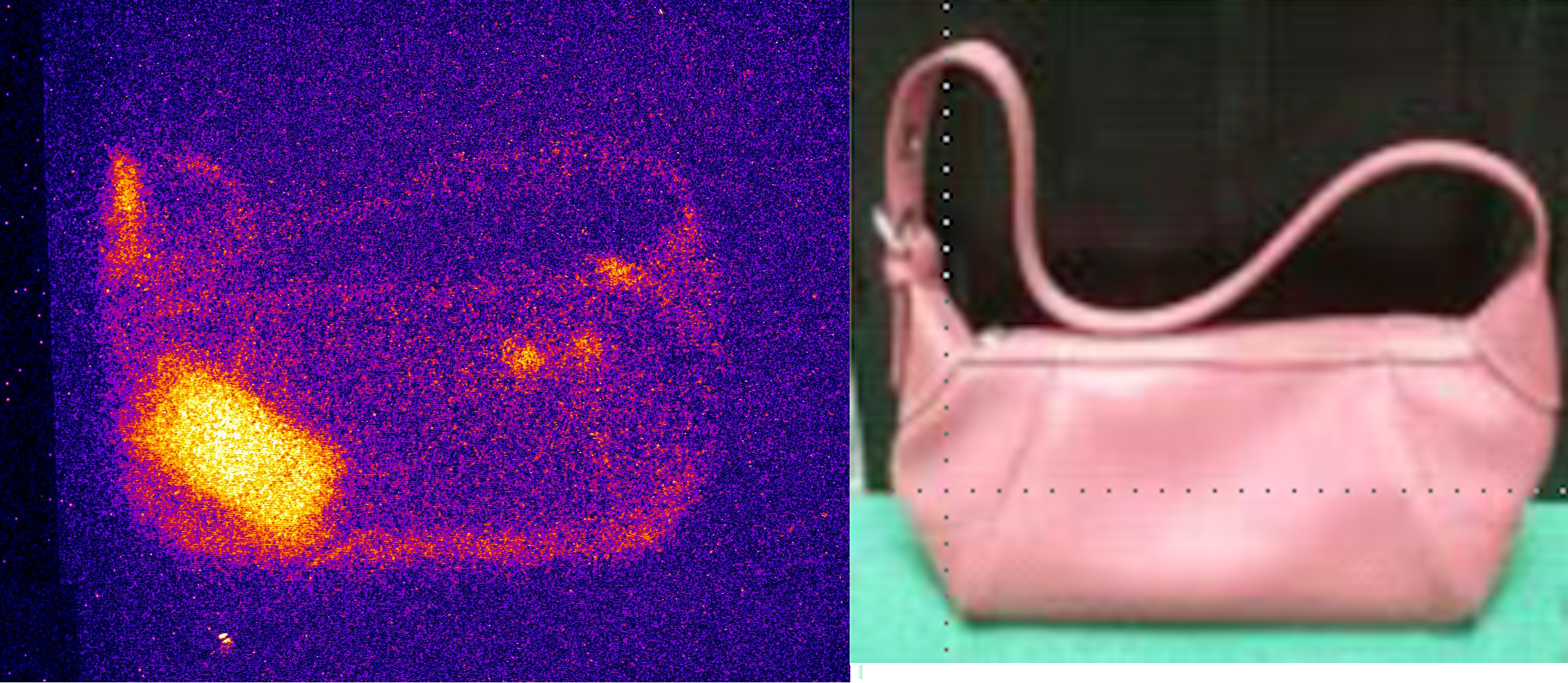
Handbag
2.45 MeV neutron imaging, handbag containing a Blackberry/phone (2008)
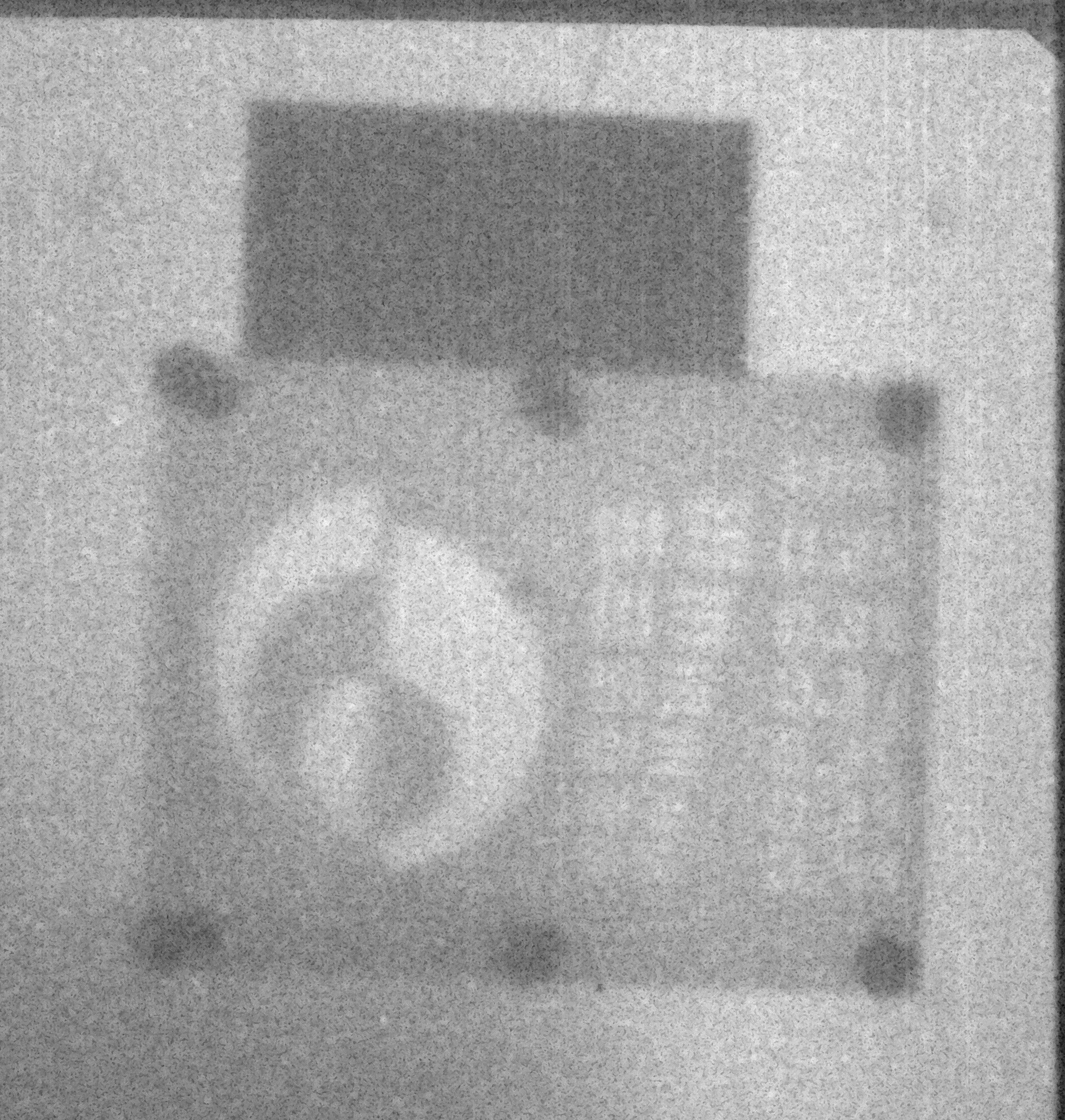
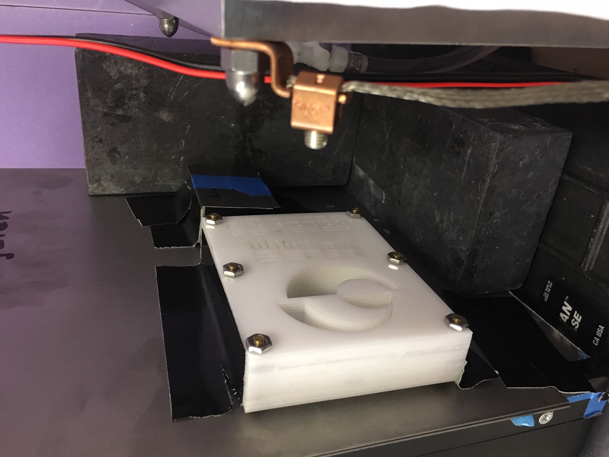
AF1951 target/standard
14.1 MeV neutron imaging, AF1951 standard machined into HDPE plastic sheets totallin 1 inch in thickness
AF1951 target/standard
14.1 MeV neutron imaging, AF1951 standard machined into HDPE plastic sheets totallin 1 inch in thickness
AF1951 target/standard
14.1 MeV neutron imaging, AF1951 standard machined into HDPE plastic sheets totallin 1 inch in thickness
AF1951 target/standard
14.1 MeV neutron imaging, AF1951 standard machined into HDPE plastic sheets totallin 1 inch in thickness
AF1951 target/standard
14.1 MeV neutron imaging, AF1951 standard machined into HDPE plastic sheets totallin 1 inch in thickness